How AI is Improving Microscopy Images with Light Tricks

Overview
- Explores the revolutionary impact of AI on microscopy image quality.
- Highlights the surprising benefits of chromatic aberration.
- Examines the exciting shift from traditional microscopy to AI-enhanced methods.
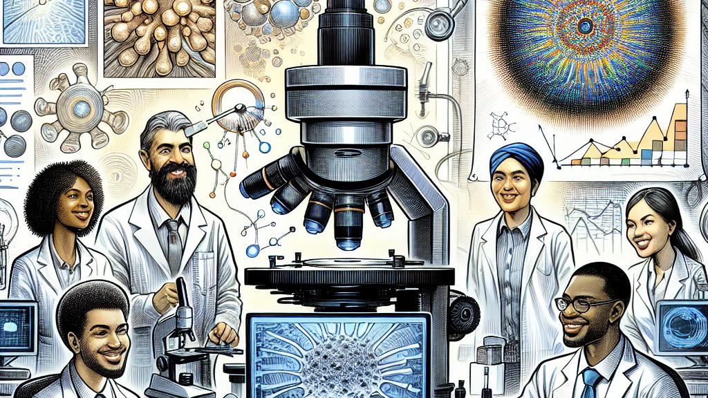
A New Era in Microscopy Powered by AI
In Germany, a team of brilliant scientists at the Center for Advanced Systems Understanding (CASUS) is transforming the field of microscopy by harnessing the potential of artificial intelligence. Historically, quantitative phase imaging (QPI) involved tedious processes requiring multiple image captures and costly equipment, which often hindered the speed and efficiency of research. However, a groundbreaking technique is emerging: by ingeniously utilizing chromatic aberration—a phenomenon where different wavelengths of light focus at different points—researchers can achieve crystal-clear imaging with just one exposure. This innovation not only accelerates research timelines but also democratizes advanced microscopy, making it more accessible for medical diagnostics and various biological applications.
Turning a Challenge into an Advantage: Chromatic Aberration
Traditionally viewed as a troublesome challenge, chromatic aberration is now seen as a valuable tool for enhancing image resolution. This clever study showcases how scientists can exploit phase shifts from red, green, and blue light using a standard RGB detector, creating an intricate through-focus stack from a single image. For instance, instead of needing to capture multiple images to analyze biological samples, researchers can streamline their workflows, thereby saving time and reducing costs. It’s truly inspiring how a previously perceived obstacle is being transformed into a vital component in the progression of microscopy techniques, pushing the scientific community towards new discoveries!
The Game-Changing Role of Generative AI in Imaging
Artificial intelligence isn’t just an addition to microscopy; it represents a remarkable shift in our approach to scientific discovery. Imagine a generative AI model, expertly trained on a massive dataset of over 1.2 million images, as it dives into the task of extracting crucial phase information from minimal input data. This game-changing ability allows researchers to glean essential cellular characteristics from just one image, enhancing both the efficiency and accuracy of their analyses. The capacity to observe and analyze cellular processes in real-time can lead to groundbreaking applications in biomedicine, radically improving our understanding of complex biological systems. Isn’t it exciting to envision the future of microscopy where these advancements will drive unprecedented discoveries and innovations?
References
Loading...