AI Method Improves Images of Thin Biological Samples

Overview
- Transformative AI technique sharpens microscopy images, revealing hidden details.
- Accessible and cost-effective, it simplifies the research process for many labs.
- Revolutionizes analysis accuracy, propelling breakthroughs in biological sciences.
Revolutionizing Microscopy
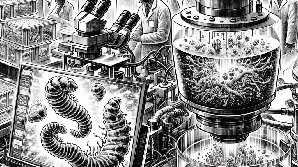
In the dynamic world of biological research in the United States, scientists at the prestigious Howard Hughes Medical Institute’s Janelia Research Campus are pioneering an innovative AI technique that is set to transform the field of microscopy. Imagine looking at a worm embryo or a slice of thick tissue: while you might start off with a clear view, the further you peer, the blurrier the images become. This blurriness can hinder scientists’ efforts to capture the delicate nuances of life at a cellular level. Traditionally, tackling this challenge required costly adaptive optics systems—a luxury that many labs simply can't afford. Fortunately, the emergence of this groundbreaking AI method provides a refreshing alternative, offering pristine images without the need for expensive upgrades or complicated apparatus.
How It Works
So, how does this remarkable technique work, you ask? The talented researchers at the Shroff Lab first devised a model to understand the specific ways in which image quality diminishes as they collect images from greater depths. By capturing clear images from the top layers of a sample and employing sophisticated deep learning algorithms, they managed to simulate the distortions that occur deeper in the sample. As a result, they trained a neural network capable of reversing these distortions, allowing them to produce high-quality images across the entire sample. What's truly impressive is that this revolutionary approach necessitates only a standard microscope and a computer with a decent graphics card, making it incredibly accessible to a plethora of biological researchers eager to enhance their imaging capabilities.
Impacts on Biological Research
The implications of this AI-enhanced imaging technique are monumental, promising to reshape the landscape of biological research. For example, researchers can now count the number of cells in developing worm embryos with stunning accuracy, a vital step in unraveling the mysteries of embryonic development. Furthermore, this method allows scientists to trace complex vascular structures within intact mouse embryos, unveiling details that could change our understanding of organ formation and function. Just picture the ability to visualize the intricate workings of mitochondria within mouse livers and hearts in vivid detail! This clarity not only elevates scientific observation but ignites the potential for groundbreaking discoveries in regenerative medicine and other critical fields. With this AI advancement, researchers are not just achieving clearer images; they are embarking on an exciting journey towards deeper biological understanding and unprecedented scientific breakthroughs.
References
Loading...